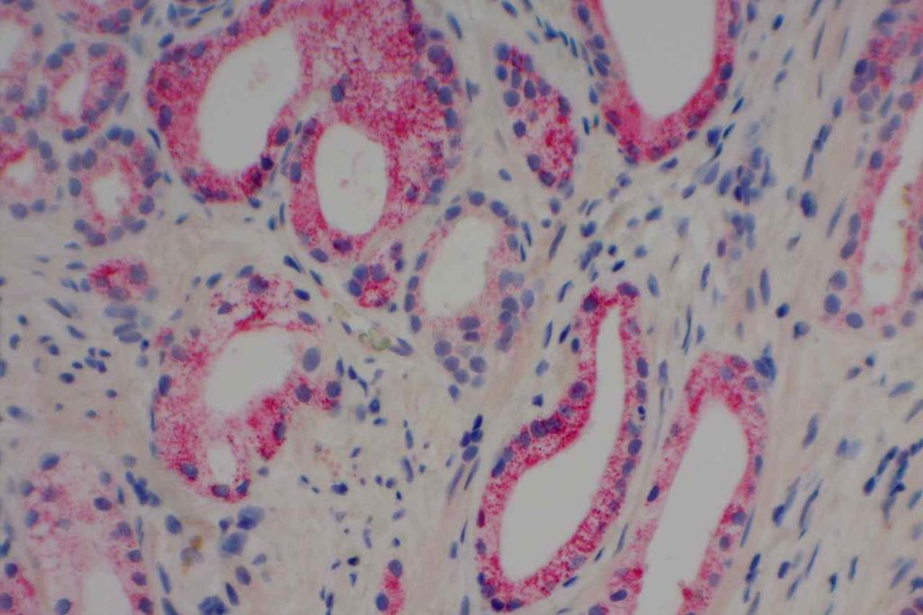
AMACR (alpha-methylacyl coenzyme) is a racemese most commonly used in the diagnosis of prostate cancer. AMACR shows excellent (though not perfect) sensitivity and specificity for prostate adenocarcinoma in prostate tissue. There are several pitfalls to be aware of with AMACR in the non-prostate gland setting. Expression is seen in normal hepatocytes, proximal renal tubule epithelium, and bronchial epithelium. Metanephric adenoma of the bladder has also been reported to be positive for AMACR.
From a renal carcinoma standpoint, AMACR is expressed in a high percentage of papillary RCCs and translocation associated RCC (TFE3) (Shen, et al., Wilkerson, et al. & Al-Ghawi et al.), but is rarely expressed by other subtypes of RCC. Many other tumor types have been shown to express AMACR (see table below).
AMACR over expression (moderate to strong staining) in various tissue sites/types (Zhou, et. al.)
|
Tumor / Tissue
|
Expression (%)
|
|
Colorectal Adenocarcinoma
|
92%
|
|
Prostate Adenocarcinoma
|
83%
|
|
Breast Carcinoma
|
44%
|
|
High Grade PIN
|
64%
|
|
Colon Adenomas
|
75%
|
|
Ovarian
|
~60%
|
|
Melanoma
|
~40%
|
|
Lung
|
~35%
|
|
Urothelial
|
~30%
|
|
Renal Cell Carcinoma
|
~30%
|
|
Lymphoma
|
~30%
|
Carcinoma in situ of the urothelium has been shown to express AMACR in 50-78% of cases by Aron, et. al. AMACR has also been shown to have increasing expression levels in Barrett’s esophagus with dysplasia and esophageal adenocarinoma. Although it is unclear if anyone uses AMACR on a routine basis in cases of Barrett’s esophagus.
Pitfalls
An important point is to understand that AMACR is very helpful (especially as part of a panel with basal markers) in differentiating benign prostate glands from prostate adenocarcinoma, but AMACR is not a specific marker of prostate gland differentiation.
Photomicrographs

References
Shen, SS. “Role of Immunohistochemistry in Diagnosing Renal Neoplams: When Is It Really Useful?”Arch Pathol Lab Med, Vol. 136, April 2012. pp. 410-417.
Jiang, Z., Li, C., Fischer, A., Dresser, K., & Woda, B. A. (2005). Using an AMACR (P504S)/34bE12/p63 Cocktail for the Detection of Small Focal Prostate Carcinoma in Needle Biopsy Specimens. American Journal of Clinical Pathology, 123(2), 231–236. doi:10.1309/1G1NK9DBGFNB792L
Yang, X. J., Wu, C.-L., Woda, B. A., Dresser, K., Tretiakova, M., Fanger, G. R., & Jiang, Z. (2002). Expression of alpha-Methylacyl-CoA racemase (P504S) in atypical adenomatous hyperplasia of the prostate. The American Journal of Surgical Pathology, 26(7), 921–925. doi:10.1097/01.PAS.0000017328.13364.17
Aron, M., Luthringer, D. J., McKenney, J. K., Hansel, D. E., Westfall, D. E., Parakh, R., et al. (2013). Utility of a Triple Antibody Cocktail Intraurothelial Neoplasm-3 (IUN-3-CK20/CD44s/p53) and α-Methylacyl-CoA Racemase (AMACR) in the Distinction of Urothelial Carcinoma In Situ (CIS) and Reactive Urothelial Atypia. The American Journal of Surgical Pathology, 37(12), 1815–1823. doi:10.1097/PAS.0000000000000114
Al-Ghawi, H., Asojo, O. A., Truong, L. D., Ro, J. Y., Ayala, A. G., & Zhai, Q. J. (2010). Application of Immunohistochemistry to the Diagnosis of Kidney Tumors. Pathology Case Reviews, 15(1), 25–34. doi:10.1097/PCR.0b013e3181d51c70
Shi, X. Y., Bhagwandeen, B., & Leong, A. S.-Y. (2008). p16, cyclin D1, Ki-67, and AMACR as markers for dysplasia in Barrett esophagus. Applied Immunohistochemistry & Molecular Morphology : AIMM / Official Publication of the Society for Applied Immunohistochemistry, 16(5), 447–452. doi:10.1097/PAI.0b013e318168598b
Wilkerson ML, Lin F, Liu H, Cheng L. The application of immunohistochemical biomarkers in urologic surgical pathology. Arch Pathol Lab Med. 2014;138(12):1643–1665. doi:10.5858/arpa.2014-0078-RA.
