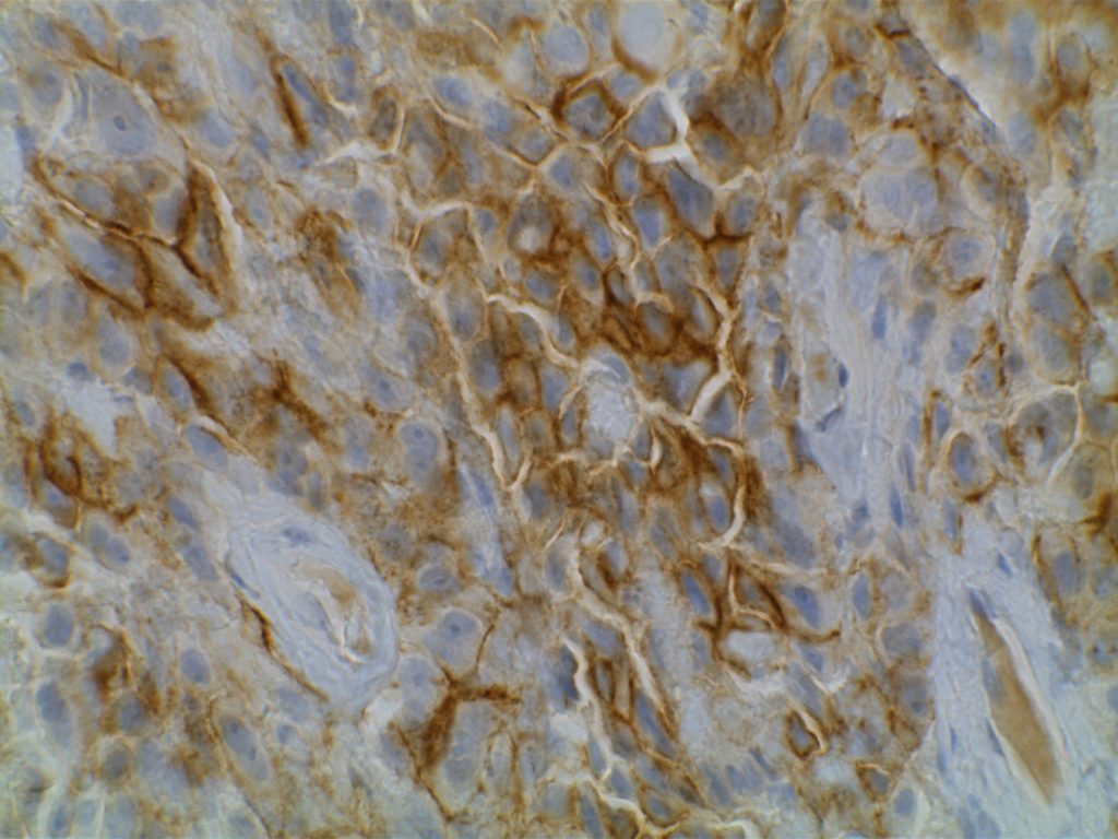
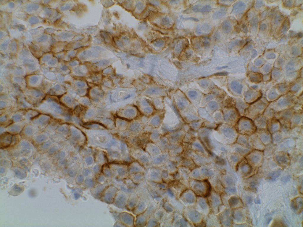
D2-40 is a marker for a sialoglycoprotein found on lymphatic endothelium. This marker is most commonly used to identify lymphatic invasion, and can help differentiate between lymphatic or vascular invasion with the additional support of a general endothelial marker (e.g. CD31 or CD34), which mark both vascular and lymphatics. It is not recommended to use D2-40 without CD31 and/or CD34 when evaluating for lymphatic invasion. Other cell types (including myoepithelial cells in the breast) can also express D2-40, which may result in misidentification of lymphatic invasion.
In addition expression is also found in germ cell neoplasia and fetal testicular monocytes. (Mohammad and Ellis)
D2-40 will also be positive in mesotheliomas, but negative in adenocarcinomas. Chu et al. showed 96% of epithelioid mesotheliomas and 65% of ovarian serous carcinomas reacted with D2-40 with membraneous staining. Other carcinomas generally did not express D2-40 in their study. There is no evidence that D2-40 is helpful with spindle-cell/sarcomatoid mesotheliomas.
Kaposi Sarcoma
D2-40 is expressed in approximately 2/3rds of cases of Kaposi’s Sarcoma. It stains in a similar pattern as CD31 and CD34.
Germ Cell Tumors
Seminomas show expression for D2-40, along with a subset of embryonic carcinomas. Yolk sac tumors are not know to express D2-40. (Bai, et al.)
Photomicrographs


References
Bai S, Wei S, Pasha TL, Yao Y, Tomaszewski JE, Bing Z. Immunohistochemical Studies of Metastatic Germ-Cell Tumors in Retroperitoneal Dissection Specimens: A Sensitive and Specific Panel. International Journal of Surgical Pathology. 2013;21: 342–351. doi:10.1177/1066896912471849
Rosado FGN, Itani DM, Coffin CM, Cates JM. Utility of immunohistochemical staining with FLI1, D2-40, CD31, and CD34 in the diagnosis of acquired immunodeficiency syndrome-related and non-acquired immunodeficiency syndrome-related Kaposi sarcoma. Arch Pathol Lab Med. 2012;136: 301–304. doi:10.5858/arpa.2011-0213-OA
Ordóñez NG. Immunohistochemical diagnosis of epithelioid mesothelioma: an update. Arch Pathol Lab Med. 2005;129: 1407–1414.
Wick MR. Immunohistochemical approaches to the diagnosis of undifferentiated malignant tumors. Annals of Diagnostic Pathology. 2008;12: 72–84. doi:10.1016/j.anndiagpath.2007.10.003
Marchevsky AM. Application of immunohistochemistry to the diagnosis of malignant mesothelioma. Arch Pathol Lab Med. 2008;132: 397–401.
Mohammed RAA, Martin SG, Gill MS, Green AR, Paish EC, Ellis IO. Improved methods of detection of lymphovascular invasion demonstrate that it is the predominant method of vascular invasion in breast cancer and has important clinical consequences. Am J Surg Pathol. 2007;31: 1825–1833. doi:10.1097/PAS.0b013e31806841f6
