General
Monoclonal and polyclonal antibodies are available to H. pylori for use in paraffin embedded tissue. While less specific, other stains (e.g. silver stains, Giemsa, modified Steiner, etc.) may be used to highlight the bacterial organisms. IHC stains provide the most sensitive and specific marker for identification. Additionally, IHC stains for H. pylori have much lower background than special stains, making it easier for the pathologist to make the diagnosis.
Stain Interpretation
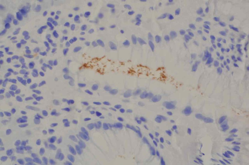
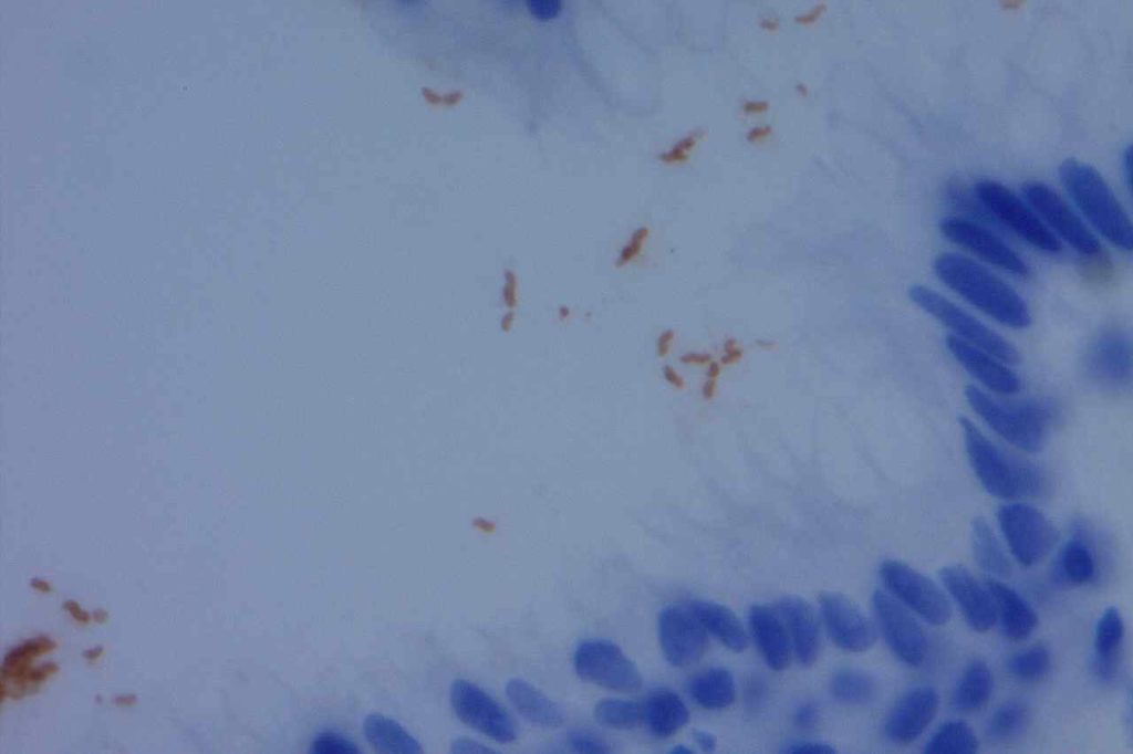
For a case to be positive, there must be staining and bacterial morphology (curvilinear organisms). There are several pitfalls to be aware of to interpret H. pylori stains:
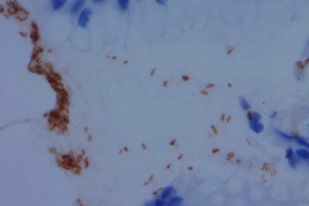
- “Junk Staining” on the surface as a result of neutral red or other pigmented stains used as the time of grossing to help visualize tissue while cutting. This most commonly occurs when stains used at the grossing station are not filtered regularly (or are just old) with subsequent precipitate material.
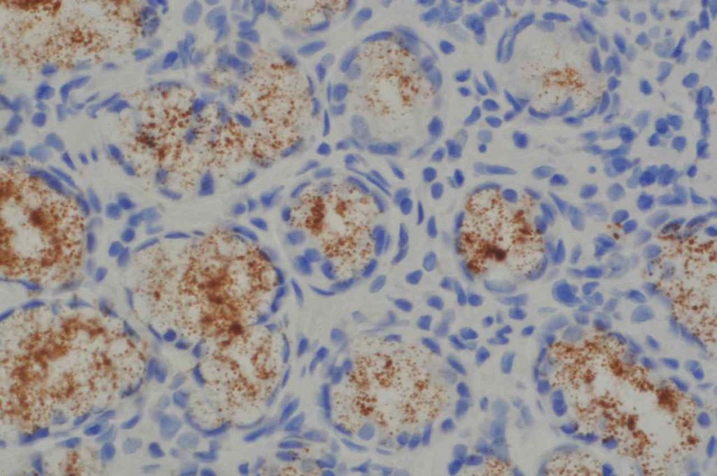
- Some antibodies (e.g. Novocastra monoclonal) may show cross-reactivity with cytoplasmic antral gland granules. This is most confusing when the granules are dislodged from the cytoplasmic location during the biopsy procedure and are distributed along the mucosal surface. They have a characteristic appearance of variably sized round granules.
Stain Sensitivity and Specificity (Hartman, et al)
|
Stain
|
Sensitivity
|
Specificity
|
|
H&E
|
83%
|
100%
|
|
Giemsa
|
62%
|
97%
|
|
Warthin-Starry
|
62%
|
98%
|
|
IHC Staining
|
97-100%
|
100%
|
General Comment
The Rodger C. Haggitt Gastrointestinal Pathology Society has published a consensus recommendation that special stains to evaluate for Helicobacter organisms are appropriate when there is chronic gastritis or chronic active gastritis without evidence of organisms on routine H&E sections. Routine performance of special stains upfront on all biopsies for Helicobacter is NOT recommended. (Batts, et al)
Please see the image example for demonstration of proper staining and artifacts.


References:
Hartman, D. J., & Owens, S. R. (2012). Are routine ancillary stains required to diagnose helicobacter infection in gastric biopsy specimens?: an institutional quality assurance review. American Journal of Clinical Pathology, 137(2), 255–260. doi:10.1309/AJCPD8FFBJ5LSLTE
Riba, A. K., Ingeneri, T. J., & Strand, C. L. (2011). Improved Histologic Identification of Helicobacter pylori by Immunohistochemistry Using a New Novocastra Monoclonal Antibody. Laboratory Medicine, 42(1), 35–39. doi:10.1309/LMAGPAENJKNARI4Z
Batts, K. P., Ketover, S., Kakar, S., Krasinskas, A. M., Mitchell, K. A., Wilcox, R., et al. (2013). Appropriate use of special stains for identifying Helicobacter pylori: Recommendations from the Rodger C. Haggitt Gastrointestinal Pathology Society. The American Journal of Surgical Pathology, 37(11), e12–22. doi:10.1097/PAS.0000000000000097
