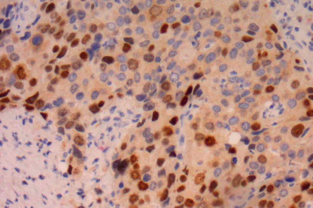
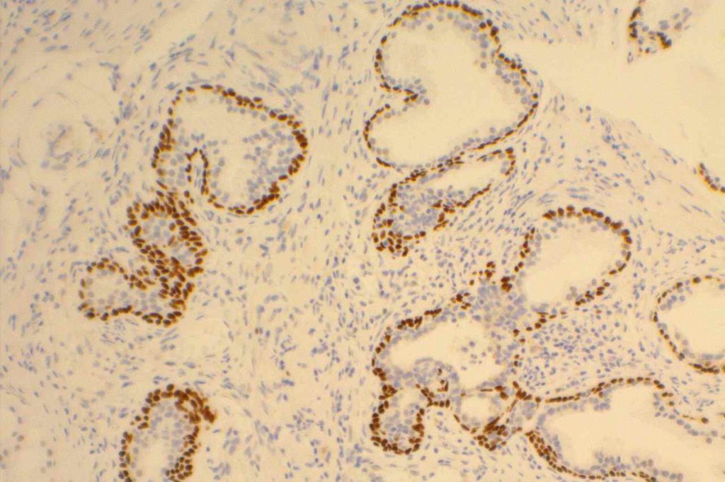
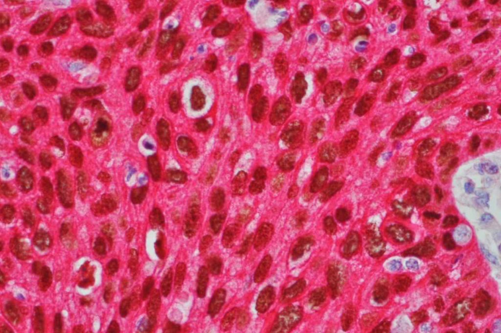
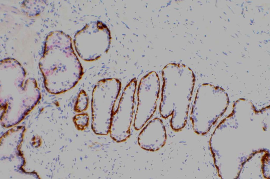
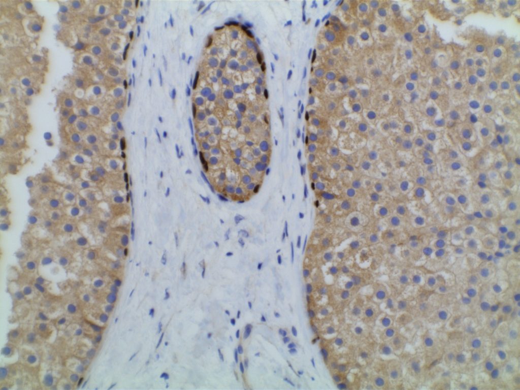
p63 is a nuclear transcription marker used in the identification of tumors subtypes and tissue structures such as: myoepithelial cell layer (breast), basal cell layer (prostate), salivary gland tumors, squamous cell carcinoma (most sensitive marker), skin appendage tumors, and urothelial carcinoma. As a general rule, transcription markers like p63 show a strong diffuse nuclear expression diagnostically.
Diagnostic Applications
|
Cell Differentiation
|
Comments
|
|
Myoepithelial Cells
|
Will stain cells in tumors of myoepithelial origin such as salivary gland tumors.
|
|
Urothelial Cells
|
70-100% of urothelial carcinomas will express p63
|
|
Squamous Cells
|
Squamous Cell Carcinomas from most locations will mark with p63 resulting in a high sensitivity.
|
|
Stromal Invasion
|
p63 can be very helpful to define stromal invasion, as it normally marks myoepithelial cells in breast tissue and basal cells in prostate tissue, which are lost in invasive carcinomas. Generally recommended to be used with a cytoplasmic myoepithelial marker in breast lesions (e.g. smooth muscle myosin or calponin).
|
p63 expression in selected tumors, may have prognostic implications for Breast, Lung, and Bladder carcinomas.
GU Pathology
p63 is expressed in a high percentage of urothelial carcinomas, but is negative in renal cell carcinomas. A small percentage of collecting duct carcinomas (14%) have been reported to express p63. p63 may be helpful in differentiation of renal urothelial carcinoma and poorly differentiated renal cell carcinoma (RCC).
Arch Pathol Lab Med-Vol.135, January, 2011
|
Tumor
|
p63 Expression (%)
|
|
Urothelial Carcinoma
|
70-100%
|
|
Renal Cell Carcinoma
|
0%
|
|
Sarcomatoid Urothelial Carcinoma
|
50%
|
|
Collecting Duct Carcinoma
|
14%
|
Placental Sit Trophoblastic Tumor
p63 is expressed in intermediate trophoblasts associated with placental site nodules, but is negative in placental site trophoblastic tumors (epithelioid trohphoblastic tumors also express p63) [Mittal, et. al.].
Undifferentiated Carcinomas
CK5/6 co-expression with p63 is a sensitive (77%) and specific (96%) marker for squamous cell carcinoma. p63 appears to be very specific for squamous and urothelial origin. Only rare other non-squamous cell carcinomas show >50% expression of p63. [Kaufman, O., et. al.]
Small Cell Carcinoma
Small cell carcinomas express p63 in approximately 77% of cases (n=14). [Au, NH, et. al.] This could be a potential pitfall if one is considering a poorly differentiated squamous cell carcinoma and not a poorly differentiated neuroendocrine carcinoma.
Microscopic Images
References
Hadi, AIMM Annual Meeting, “The Thirty Most Important Antibodies”, presentation, 2011.
Truong, LD, et. al. “Immunohistochemical Diagnosis of Renal Neoplasms.” Archives of Pathology and Laboratory Medicine. 2011;135:92-109.
Mittal, K, et. al. “Aplplication of Immunohistochemistry to Gynecologic Pathology.” Archives of Pathology and Laboratory Medicine. Vol. 132, March 2008.
Kaufmann, O., et. al. “Value of p63 and CK 5/6 as IHC Markers for the Differential Dx. of Poorly Differentiated and Undifferentiated Carcinomas.” American Journal of Clinical Pathology. 2001;116:823-830
Au NHC, Gown AM, Cheang M, et al. P63 expression in lung carcinoma: a tissue microarray study of 408 cases. Appl Immunohistochem Mol Morphol. 2004;12(3):240–247.
Zhao L, Yang X, Khan A, Kandil D. Diagnostic role of immunohistochemistry in the evaluation of breast pathology specimens. Arch Pathol Lab Med. 2014;138: 16–24. doi:10.5858/arpa.2012-0440-RA
