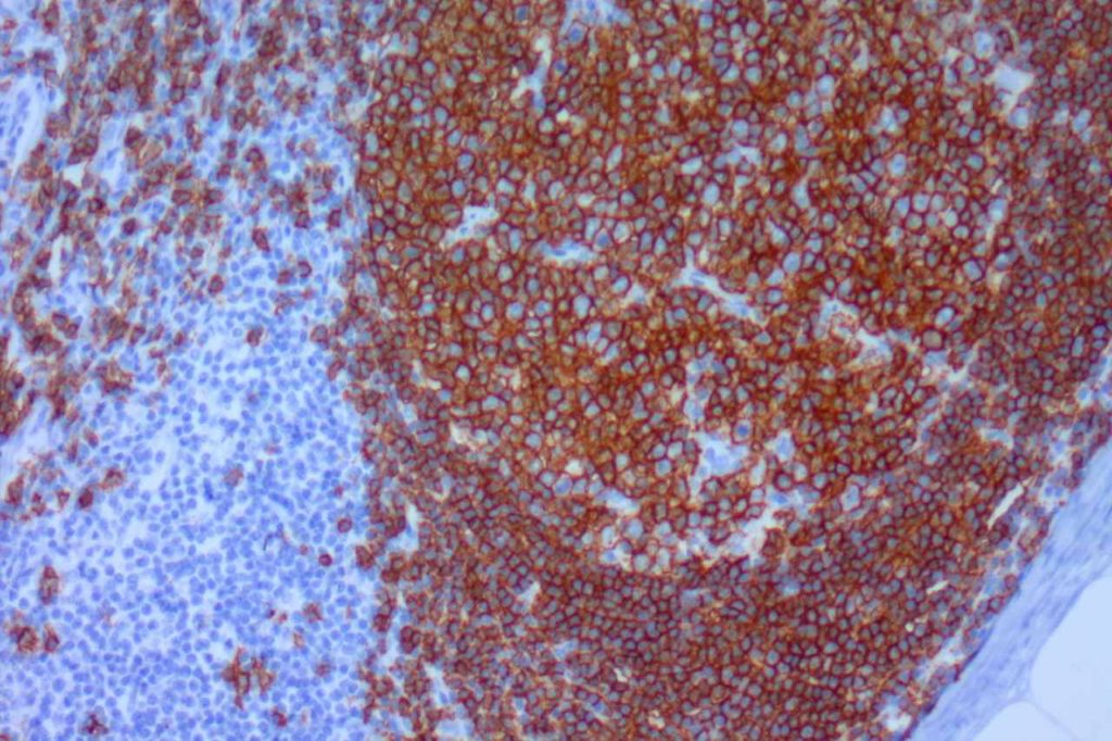
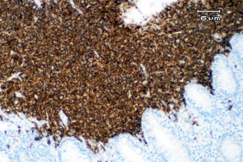
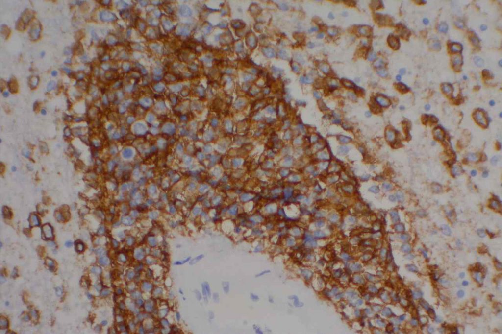
CD20 is a pan B-cell marker and is one of the most utilized hematopathology stains. The staining pattern morphology is membraneous and cytoplasmic. The sensitivity and specificity for mature B-cell neoplasms is very high.
An important caveat is that up to 27% of B cell neoplasms treated with rituximab will lose/significantly reduce CD20 expression at some point in the disease process. There is also evidence that other B-cell markers may be down regulated in response to rituximab therapy (e.g. PAX-5, etc.), and a panel of B-cell markers should be considered to fully evaluate for loss of CD20 expression. If a B-cell lymphoma loses CD20 expression, it may have a decreased response rate to rituximab therapy (from 70% to 25%). A subset of cases may recover CD20 expression over time, and expression in future biopsies should be studied and commented on.
Alternative B-cell markers including CD19, PAX-5 and CD79a can be used in comparison with CD20 to determine if and how much loss of CD20 expression has occured.
A small subset of abnormal (~20%) and benign plasma cells and Hodgkin cells (~20%) may express CD20.
CD20 Expression
- Pan B-cell Marker
- Mature B-cells
- B-cell Lymphomas
- Hematogones
- Plasma cells (small subset)
- B-cell Acute Lymphoblastic Lymphoma (~20%)
General pathology practice note: As a matter of routine staining and evaluation of lymphoid populations, CD3 and CD20 serve as a starting point and X-Y axis for the interpretation of all other IHC markers.
Photomicrographs


References
Bone Marrow IHC. Torlakovic, EE, et. al. American Society for Clinical Pathology Pathology Press © 2009. pp. 57.
Chu, P. G., Loera, S., Huang, Q., & Weiss, L. M. (2006). Lineage Determination of CD20- B-Cell Neoplasms: An Immunohistochemical Study. American Journal of Clinical Pathology, 126(4), 534–544. doi:10.1309/3WG32YRAMQ7RB9D4
Maeshima AM, Taniguchi H, Fukuhara S, Morikawa N, Munakata W, Maruyama D, et al. Follow-up data of 10 patients with B-cell non-Hodgkin lymphoma with a CD20-negative phenotypic change after rituximab-containing therapy. Am J Surg Pathol. 2013;37: 563–570. doi:10.1097/PAS.0b013e3182759008
Boyd SD, Natkunam Y, Allen JR, Warnke RA. Selective immunophenotyping for diagnosis of B-cell neoplasms: immunohistochemistry and flow cytometry strategies and results. Appl Immunohistochem Mol Morphol. 2013;21: 116–131. doi:10.1097/PAI.0b013e31825d550a
