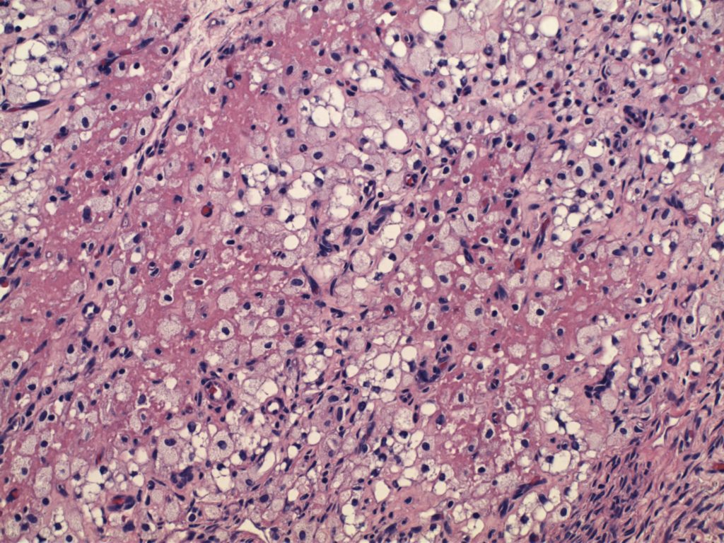
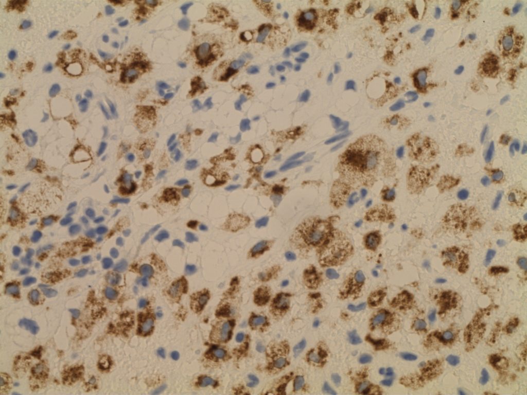
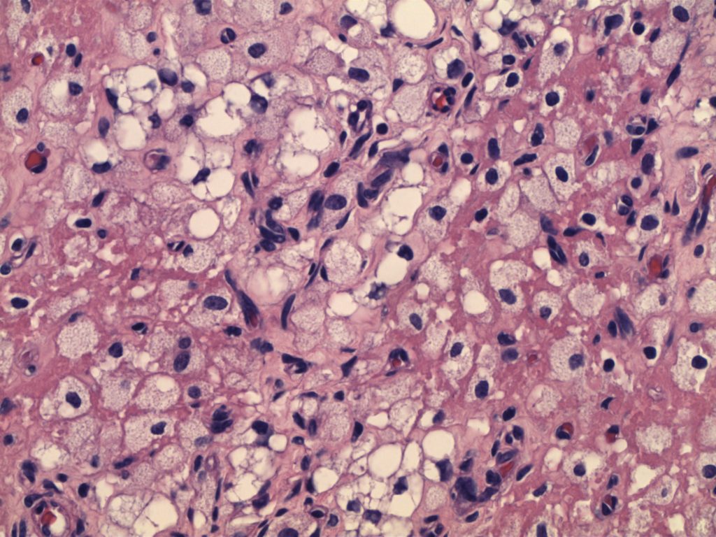
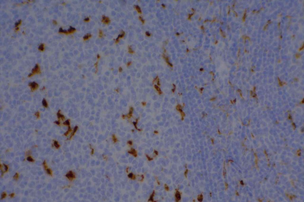
CD68 (gp110) is a glycosylated lysosomal transmembrane glycoprotein, which is normally expressed on monocytes, macrophages, dendritic cells, neutrophils, basophils, myeloid progenitor cells, mast cells, activated T-cells, and a subset of blasts. The staining pattern is generally cytoplasmic. Almost all MPO+ bone marrow cells will express CD68.
In the setting of leukemia cutis, CD68 was expressed in >95% of cases (Benet, et. al). Typically, CD68 is used to identify histiocytes. Unfortunately, CD68 lacks specificity and the morphologic differential diagnosis overlaps in many situations, and use as part of a larger panel yields more specific results.
CD68R (PGM-1) is one of the epitopes of CD68, and appears to be the most monocyte specific marker of the CD68 family. Expression in monocytic differentiated AMLs is reported to be 92-94% (Rollins-Raval, et. al)
Photomicrographs


References
Bone Marrow IHC. Torlakovic, EE, et. al. American Society for Clinical Pathology Pathology Press © 2009. pp. 114.
Bénet C, Gomez A, Aguilar C, Delattre C, Vergier B, Beylot-Barry M, et al. Histologic and immunohistologic characterization of skin localization of myeloid disorders: a study of 173 cases. Am J Clin Pathol. 2011;135: 278–290. doi:10.1309/AJCPFMNYCVPDEND0
Cronin DMP, George TI, Sundram UN. An updated approach to the diagnosis of myeloid leukemia cutis. Am J Clin Pathol. 2009;132: 101–110. doi:10.1309/AJCP6GR8BDEXPKHR
Rollins-Raval MA, Roth CG. The value of immunohistochemistry for CD14, CD123, CD33, myeloperoxidase and CD68R in the diagnosis of acute and chronic myelomonocytic leukaemias. Histopathology. 2012;60: 933–942. doi:10.1111/j.1365-2559.2012.04175.x
