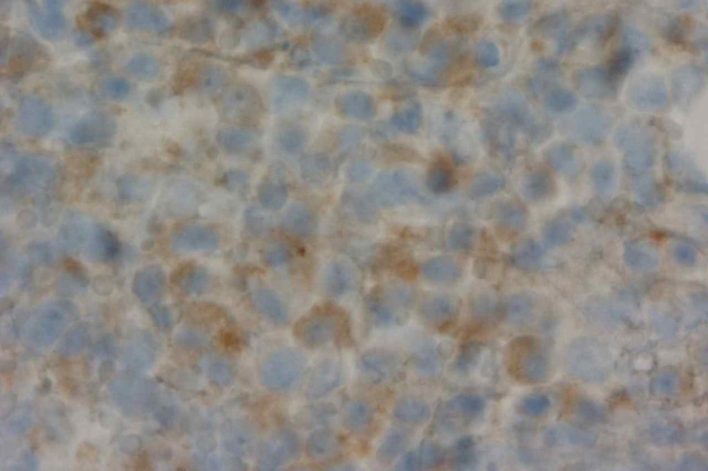
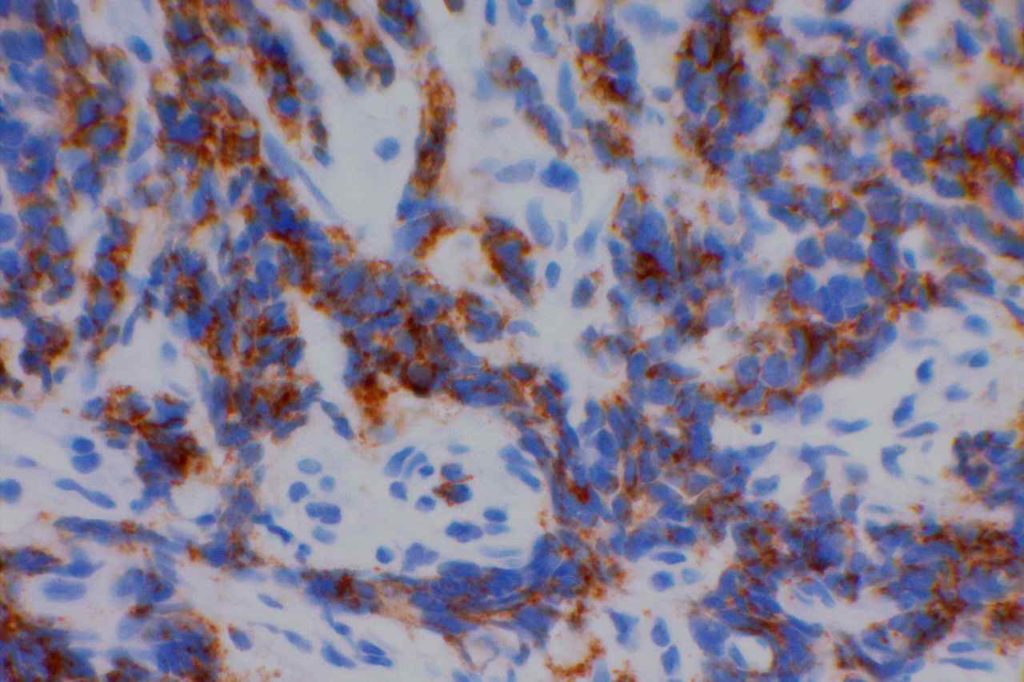
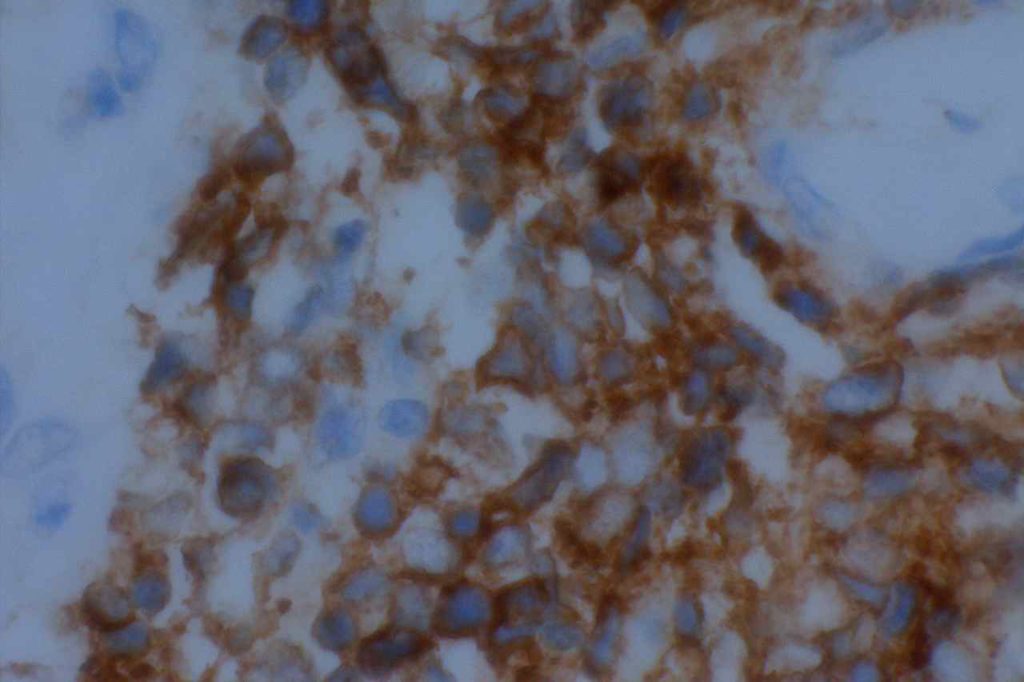
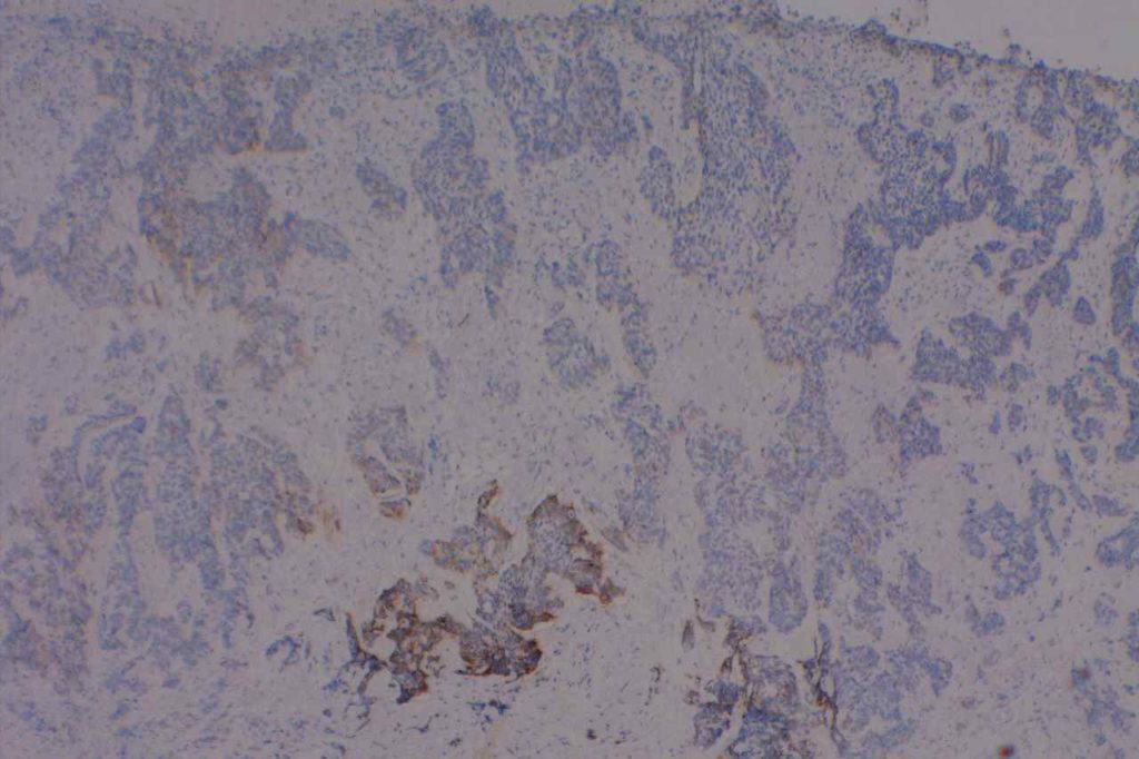
CD56 (NCAM – neural-cell adhesion molecule) is expressed on the surface of neuroendocrine epithelial cells, some Schwann cells, and some neuroendocrine tumors. In undifferentiated tumors, CD56 can be used as a screening marker for neuroendocrine differentiation. It appears to be more sensitive than synaptophysin or chromogranin A in most situations, but Ishida, et. al found CD56 to be the least sensitive of the three in neuroendocrine carcinomas of the stomach (47% compared to 94% and 86% for synaptophysin and chromogranin A, respectively).
CD56 also marks a subset of hematopoeitic and gonadal-stromal cells. Expression of CD56 alone should does not have significant specificity.
CD56 Sensitivity for high grade neuroendocrine tumors
- Stomach – 47% (Ishida, et. al)
- Esophagus – 93% (Huang, et. al)
- Lung – >90% (Travis, et. al)
Hematopathology
- Marks approximately ~20-50% of cases of leukemia cutis.
- Plasma cell myeloma – Abnormal plasma cells will often express CD56 in addition to CD138, and can be very helpful when flow cytometry and kappa/lambda studies fail to identify an abnormal plasma cell population.
- Acute Myeloid Leukemia (~13%; 38% in monoblastic AML)
- Multiple Myeloma (>70%)
- MGUS (<10%)
- CD4+CD56+ Plasmacytoid Dendritic Cell Tumor (Hematodermic Neoplasm)
- NK & T-cell Neoplasms (Subset)
- Hepatosplenic T-cell Lymphoma (CD56+, CD57=)
- NK Cells
Other
Basal cell carcinomas (BCC) show strong membrane and less intense cytoplasmic staining with CD56. Important not to use with the differential diagnosis of merkel cell carcinoma. Squamous cell carcinomas (SCC) do not express CD56, and in the BCC vs. SCC differential CD56 may be helpful.
CD56 Expression Pattern – Other
- BM Osteoblasts
- Neuroendocrine Neoplasms
- Basal cell carcinoma
Photomicrographs


References
Cronin DMP, George TI, Sundram UN. An updated approach to the diagnosis of myeloid leukemia cutis. Am J Clin Pathol. 2009;132: 101–110. doi:10.1309/AJCP6GR8BDEXPKHR
Wick MR. Immunohistochemical approaches to the diagnosis of undifferentiated malignant tumors. Annals of Diagnostic Pathology. 2008;12: 72–84. doi:10.1016/j.anndiagpath.2007.10.003
Bone Marrow IHC. Torlakovic, EE, et. al. American Society for Clinical Pathology Pathology Press © 2009. pp. 104.
Ishida M, Sekine S, Fukagawa T, Ohashi M, Morita S, Taniguchi H, et al. Neuroendocrine carcinoma of the stomach: morphologic and immunohistochemical characteristics and prognosis. Am J Surg Pathol. 2013;37: 949–959. doi:10.1097/PAS.0b013e31828ff59d
Huang Q, Wu H, Nie L, Shi J, Lebenthal A, Chen J, et al. Primary high-grade neuroendocrine carcinoma of the esophagus: a clinicopathologic and immunohistochemical study of 42 resection cases. Am J Surg Pathol. 2013;37: 467–483. doi:10.1097/PAS.0b013e31826d2639
Cronin DMP, George TI, Reichard KK, Sundram UN. Immunophenotypic analysis of myeloperoxidase-negative leukemia cutis and blastic plasmacytoid dendritic cell neoplasm. Am J Clin Pathol. 2012;137: 367–376. doi:10.1309/AJCP9IS9KFSVWKGH
Bénet C, Gomez A, Aguilar C, Delattre C, Vergier B, Beylot-Barry M, et al. Histologic and immunohistologic characterization of skin localization of myeloid disorders: a study of 173 cases. Am J Clin Pathol. 2011;135: 278–290. doi:10.1309/AJCPFMNYCVPDEND0
Joshi R, Horncastle D, Elderfield K, Lampert I, Rahemtulla A, Naresh KN. Bone marrow trephine combined with immunohistochemistry is superior to bone marrow aspirate in follow-up of myeloma patients. J Clin Pathol. 2008;61: 213–216. doi:10.1136/jcp.2007.049130
Seegmiller AC, Xu Y, McKenna RW, Karandikar NJ. Immunophenotypic differentiation between neoplastic plasma cells in mature B-cell lymphoma vs plasma cell myeloma. Am J Clin Pathol. 2007;127: 176–181. doi:10.1309/5EL22BH45PHUPM8P
BELJAARDS RC, KIRTSCHIG G, BOORSMA DM. Expression of neural cell adhesion molecule (CD56) in basal and squamous cell carcinoma. Dermatol Surg. 2008;34: 1577–1579. doi:10.1111/j.1524-4725.2008.34327.x
Herling M, Jones D. CD4+/CD56+ hematodermic tumor: the features of an evolving entity and its relationship to dendritic cells. Am J Clin Pathol. 2007;127: 687–700. doi:10.1309/FY6PK436NBK0RYD4
Travis WD. Update on small cell carcinoma and its differentiation from squamous cell carcinoma and other non-small cell carcinomas. Mod Pathol. Nature Publishing Group; 2012;25: S18–S30. doi:10.1038/modpathol.2011.150
