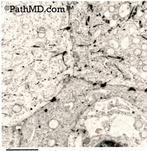
PathMD Quizes, Anatomic Electron Microscopy – 01 October 29, 2017 peferguson Make sure to subscribe to PathMD to stay up to date with new content and features!! 1. EM of a CNS tumor shows prominent interdigitating processes without a basal lamina. There are well formed desmosome junctions and prominent cytoplasmic inclusions. These findings are most consistent with:CarcinomaLymphomaSchwannomaEpendymomaMeningioma 2. EM of an undifferentiated tumor is noted to have long thin microvilli (height to width ~ 10:1). Which of the following tumors is this most consistent with:Prostate carcinoma MesotheliomaBreast adenocarcinoma Thyroid follicular carcinoma Pulmonary adenocarcinoma 3. Based on the findings in the image below for Case #5, which of the following is the best diagnosis? Electron Microscopy Set 1 Case 5MelanomaRenal Cell Carcinoma, chromophobe type Renal Cell Carcinoma, clear cell typeOncocytomaGranular Cell Tumor 4. A biopsy from the tongue shows pseudoepitheliomatous hyperplasia with histiocyte like cells underneath, which contain a granular cytoplasm. A diagnosis is suspected and confirmed with S-100 positivity. Which of the following is true with regards to this tumor:EM will show numerous premelanosomes EM will show abundant mitochondria EM will show cytoplasmic microvilli EM will show numerous lysosomesEM will show abundant rough endoplasmic reticulum 5. An undifferentiated tumor shows prominent profiles of rough endoplasmic reticulum. Which of the following markers would also be positive in this lesion?S-100 CD45Hale’s colloidal ironSynaptophysinCD138 6. EM of a CNS tumor showed intracellular lumens lined by microvilli with specialized junctions between cells and glial features. This is most conisistant with which of the following tumors:AstrocytomaEpendymomaChoroid plexus tumorCarcinomaSchwannoma 7. EM of a tumor shows rhomboid crystalline inclusions. Cytogenetics also showed a t(X;17). These findings are consistent with which of the following neoplasms?Alveolar soft part sarcoma MedulloblastomaClear cell sarcomaNeurocytomaPapillary ependymoma 8. Based on the findings in the images below for Case #3, which of the following is the best diagnosis? Electron Microscopy Set 1 Case 3 Electron Microscopy Set 1 Case 3MelanomaAdenocarcinomaSchwannomaSquamous Cell Carcinoma Mesothelioma 9. Based on the findings in the images below for Case #2, which of the following is the best diagnosis? Electron Microscopy Set 1 Case 2 Electron Microscopy Set 1 Case 2SchwannomaAdenocarcinomaGranular Cell Tumor PlasmacytomaMelanoma 10. Based on the findings in the images below for Case 1, which of the following is the best diagnosis? Electron Microscopy Set 1 Case 1 Electron Microscopy Set 1 Case 1 Electron Microscopy Set 1 Case 1MesotheliomaSquamous Cell Carcinoma AdenocarcinomaSchwannomaMelanoma 11. A specimen being prepared for electron microscopy should be fixed in:B5Bouin’sGluteraldehydeMercury based fixativeZinc formalin 12. Based on the findings in the images below for Case #4, which of the following is the best diagnosis? Electron Microscopy Set 1 Case 4 Electron Microscopy Set 1 Case 4LymphomaMeningiomaAdenocarcinomaSchwannomaSquamous Cell Carcinoma 13. EM of an undifferentiated tumor shows microvilli with long filamentous core rootlets (electron dense). This finding suggests which diagnosis:Breast adenocarcinoma GI adenocarcinoma Thyroid follicular carcinoma MesotheliomaProstate carcinoma Loading... Share this:TwitterWhatsAppFacebookEmailPrintLinkedIn Related
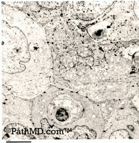 Electron Microscopy Set 1 Case 3
 Electron Microscopy Set 1 Case 3
