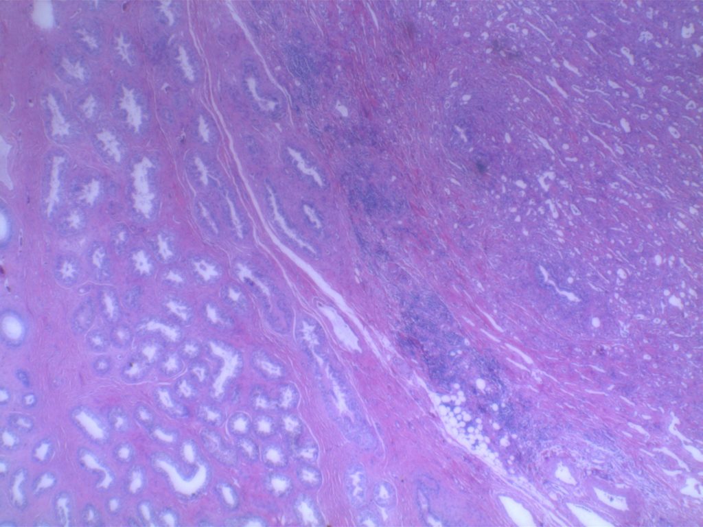
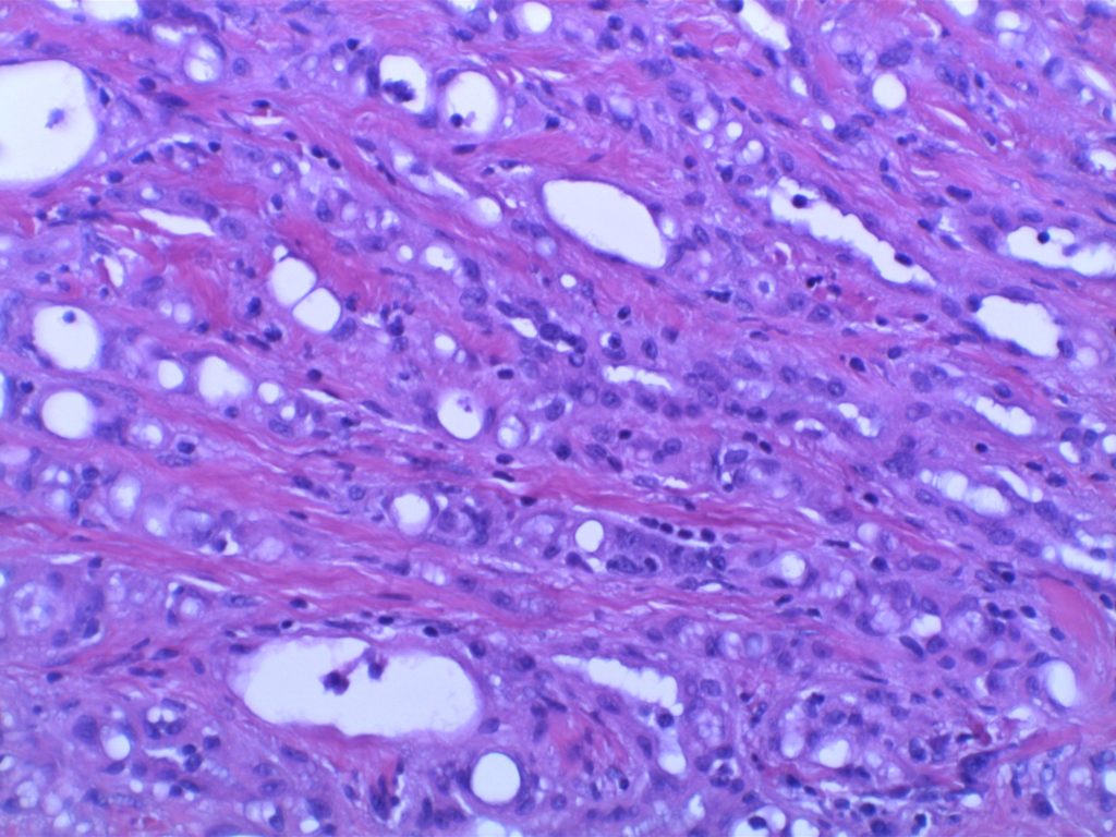
Adenomatoid tumor of the epididymis is the second most common tumor in the paratesticular region (spermatic cord lipoma is most common). These benign lesions are mesothelial in origin (usually <2 cm, but can be up to 3-5 cm), but can have significant variation in appearance which can often be “malignant” looking (cords, nests, or tubule formation). Not infrequently the pathologist may be concerned about a carcinoma.
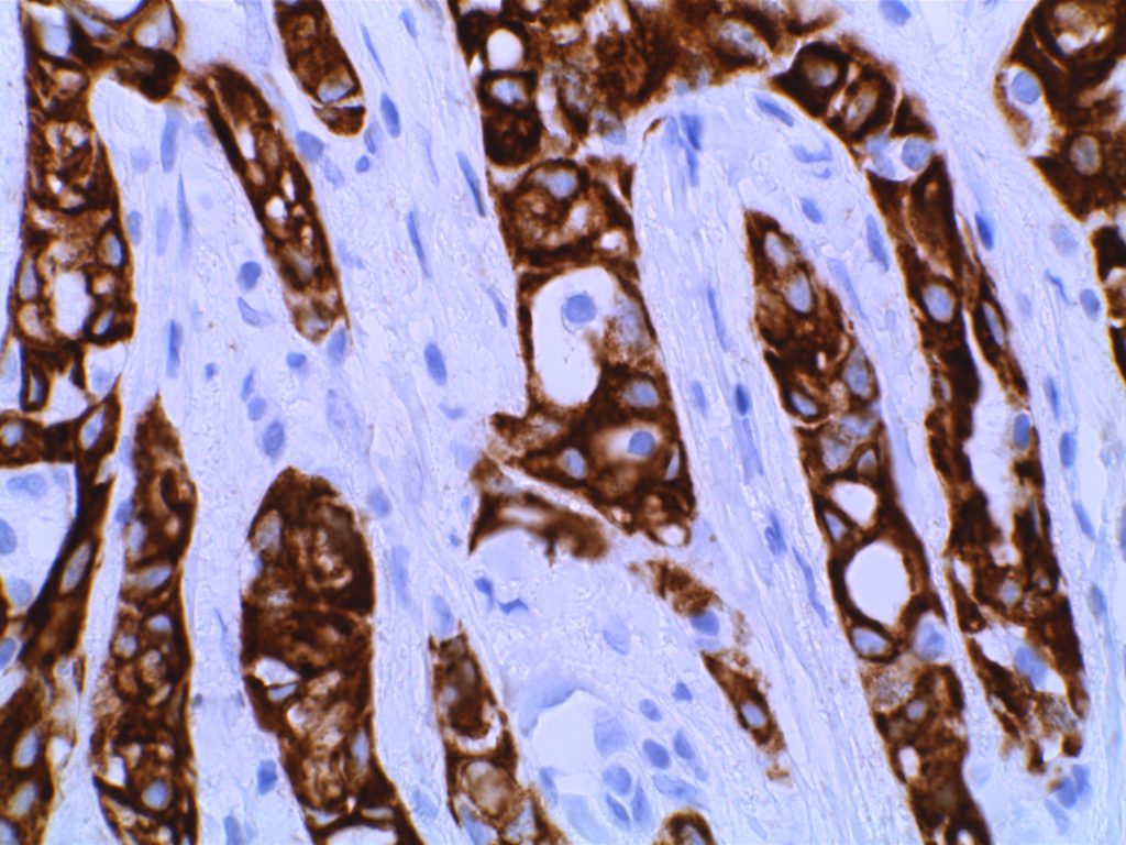
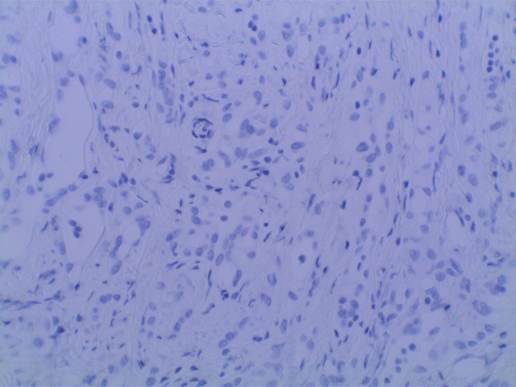
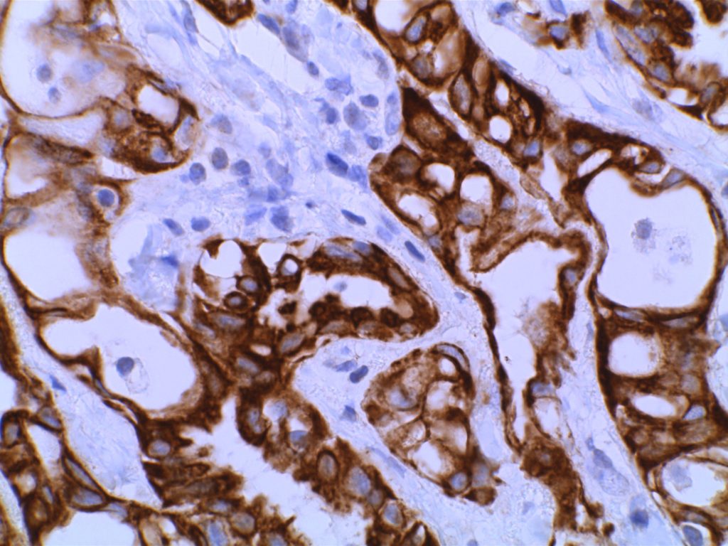
Immunohistochemistry – These lesions will express mesothelial markers, including pancytokeratin, CK7, D2-40, WT-1, calretinin, and EMA. Endothelial markers (CD34 and CD31) are not expressed, nor are other markers typically expressed in adenocarcinomas (e.g. CEA, BerEP4, MOC-31).
Photomicrographs
References
Arias-Stella JA, Williamson SR. Updates in Benign Lesions of the Genitourinary Tract. Surg Pathol Clin. 2015;8: 755–787. doi:10.1016/j.path.2015.09.001
Sternberg’s diagnostic surgical pathology.-5th ed. / senior editor, Stacey E. Mills; editors Daryl Carter … [et al.]. pp. 1990-91.
