Many different markers can be used in neoplastic lung, but the most common involve differentiation between squamous and adenocarcinoma and identifying evidence of neuroendocrine differentiation.
Squamous Versus Adenocarcinoma
Neuroendocrine Differentiation
|
(% +)
|
(% +)
|
|
|
Lung Tumors
|
|
|
| Adneocarcinoma |
79/95 (83%)
|
69/95 (73%)
|
|
42/47 (89%)
|
38/47 (81%)
|
|
27/32 (84%)
|
24/32 (75%)
|
|
11/16 (69%)
|
7/16 (44%)
|
|
0/46 (0%)
|
0/48 (0%)
|
|
3/9 (33%)
|
4/9 (44%)
|
|
0/3 (0%)
|
1/3 (33%)
|
|
Atypical Carcinoid Tumor
|
0/1 (0%)
|
1/1 (100%)
|
|
Typical Carcinoid Tumor
|
0/2 (0%)
|
1/2 (50%)
|
|
Non-pulmonary Adenocarcinomas
|
|
|
|
Colon Adenocarcinoma
|
0/5 (0%)
|
0/5 (0%)
|
|
Pancreas Adenocarcinoma
|
0/31 (0%)
|
0/31 (0%)
|
|
Breast Adenocarcinoma
|
0/17 (0%)
|
0/17 (0%)
|
|
Mesothelioma (all types)
|
0/38 (0%)
|
0/38 (0%)
|
|
Renal Cell Carcinomas
|
|
|
|
Clear Cell
|
14/41 (34%)
|
0/41 (0%)
|
|
Papillary
|
34/43 (79%)
|
0/43 (0%)
|
|
Chromophobe
|
1/34 (3%)
|
0/34 (0%)
|
|
Thyroid Lesions
|
|
|
|
Papillary Carcinoma
|
2/38 (5%)
|
37/38 (97%)
|
|
Follicular Carcinoma
|
0/15 (0%)
|
15/15 (100%)
|
|
Follicular Adenoma
|
0/28 (0%)
|
28/28 (100%)
|
CK5
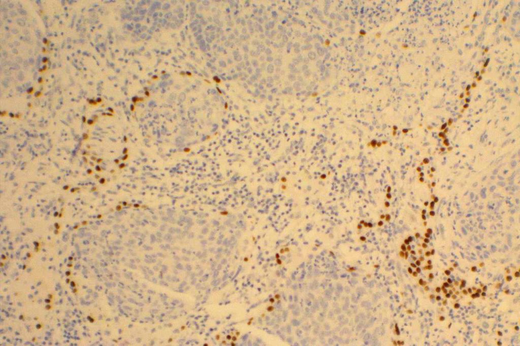
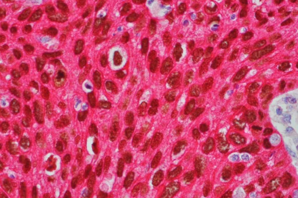
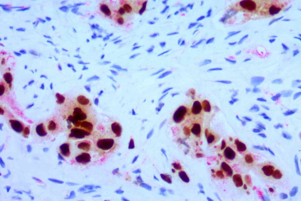
CK5 will stain approximately 70-80% of squamous cell carcinomas with a cytoplasmic staining pattern. p63 is considered slightly more sensitive, but may give varying positivity in cases of lung adenocarcinoma that can be confusing. Therefore, CK5 and p63 are often used as part of a panel to diagnose squamous cell carcinoma. Since p63 is a nuclear marker, these two stains can be performed as a double stain on a single slide with one or two different chromogens.
p63
Double Stains
Double stains can be helpful in conserving tissue for small biopsy samples, which is critical to be able to perform testing for ALK, ROS-1, and EGFR mutations. Nuclear and cytoplasmic markers paired together with different colored chromagens are preferred (e.g. TTF-1/Napsin A for adenocarcinomas and CK5/p63 for squamous cell carcinomas)
Photomicrographs


References
Bishop, J. A., Sharma, R., & Illei, P. B. (2010). Napsin A and thyroid transcription factor-1 expression in carcinomas of the lung, breast, pancreas, colon, kidney, thyroid, and malignant mesothelioma. Human Pathology, 41(1), 20–25. doi:10.1016/j.humpath.2009.06.014
